- Plot No. 361/3074 & Plot No 437/3134, Patrapada, BBSR
- +91 9040017001
- +91 8630400500

Spine MRI: A Complete Guide to Spinal Imaging and Diagnosis
Persistent back pain, neck discomfort, numbness, or tingling in the arms and legs can sometimes indicate an underlying spinal problem. While these symptoms may result from muscle strain or other common conditions, they can also be associated with disc problems, nerve compression, or changes in the spinal cord.
A Spine MRI is an advanced imaging test that helps doctors examine the structures of the spine in detail. It uses strong magnetic fields and radio waves to produce detailed images of the spinal cord, vertebrae, discs, nerves, and surrounding soft tissues.
According to a systematic review and meta-analysis published in 2022, the pooled point
prevalence of low back pain in India was 48%. The study included 97 research studies and also reported an annual prevalence of 51% and a lifetime prevalence of 66%. These findings highlight the significant burden of back pain in the Indian population.
Understanding when a Spine MRI is needed, what it can detect, and how to prepare for the scan can help patients make informed decisions about their diagnostic care.
What Is a Spine MRI?
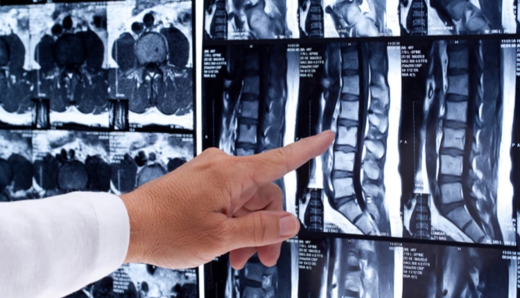
A Spine MRI (Magnetic Resonance Imaging) is a non-invasive diagnostic imaging procedure used to examine the bones, discs, nerves, spinal cord, and soft tissues surrounding the spine.
Unlike X-rays and CT scans, MRI does not use ionising radiation. Instead, it uses magnetic fields and radio waves to create detailed images of internal body structures.
Doctors may recommend a Spine MRI to investigate persistent pain, suspected nerve compression, spinal injuries, infections, tumours, or other abnormalities affecting the spinal column.
The scan helps identify structural changes and provides important information that doctors can use alongside symptoms, physical examinations, and medical history to plan appropriate treatment.
Why Is a Spine MRI Recommended?
A doctor may recommend a Spine MRI when symptoms suggest a possible problem involving the spinal cord, discs, nerves, or surrounding tissues.
Some common reasons include:
- Persistent back or neck pain: When discomfort continues or worsens despite appropriate initial treatment.
- Numbness or tingling: To investigate possible nerve irritation or compression.
- Muscle weakness: When weakness in the arms or legs may be related to spinal nerve involvement.
- Suspected slipped disc: To examine disc bulging, herniation, or related nerve compression.
- Spinal injuries: To assess damage to the spinal cord, discs, ligaments, or surrounding soft tissues.
- Unexplained neurological symptoms: To investigate symptoms that may involve the spinal cord or nerve roots.
- Follow-up after surgery: To evaluate the spine when new or recurring symptoms develop.
An MRI is not necessary for every episode of back pain. Doctors generally recommend imaging when the patient’s symptoms, examination findings, or medical history indicate that further investigation is appropriate.
What Conditions Can a Spine MRI Detect?
A Spine MRI provides detailed images that help doctors investigate several spinal and neurological conditions.
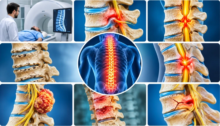
1. Herniated or Slipped Disc
A herniated disc occurs when the soft inner material of an intervertebral disc pushes through its outer layer. It may irritate or compress nearby nerves, causing pain, tingling, numbness, or weakness.
An MRI helps identify the location and extent of disc herniation and whether nearby nerves are affected.
2. Spinal Stenosis
Spinal stenosis refers to the narrowing of spaces within the spinal canal or nerve passageways. This narrowing can put pressure on the spinal cord or spinal nerves.
A Spine MRI helps doctors examine the narrowed areas and assess possible nerve compression.
3. Degenerative Disc Disease
As people age, spinal discs may gradually lose water content and change in structure. These changes can contribute to stiffness, discomfort, and other spinal symptoms.
MRI helps evaluate disc degeneration and associated structural changes.
4. Spinal Cord Compression
Spinal cord compression occurs when pressure is placed on the spinal cord due to disc herniation, spinal stenosis, injuries, tumours, or other abnormalities.
An MRI provides detailed images of the spinal cord and surrounding structures, helping doctors assess the cause and extent of compression.
5. Spinal Tumours
Abnormal growths can develop within or around the spinal column and spinal cord. A Spine MRI can help identify suspicious masses, determine their location, and assess their relationship with surrounding structures.
6. Spinal Infections
Infections involving the vertebrae, intervertebral discs, or surrounding tissues may cause pain and other symptoms.
MRI can help detect changes associated with conditions such as discitis, vertebral osteomyelitis, and spinal epidural infections.
7. Spinal Injuries
Following an accident or injury, an MRI may help assess damage to the spinal cord, discs, ligaments, and soft tissues. It is particularly useful when doctors need detailed information about structures that may not be clearly visible on an X-ray.
What Are the Different Types of Spine MRI?
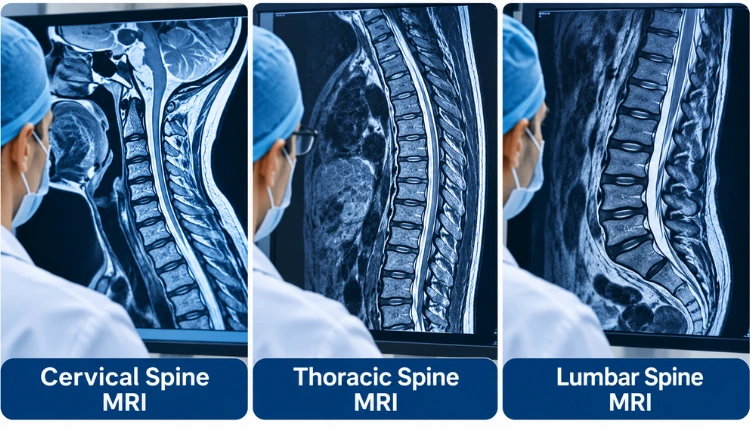
The spine is divided into different anatomical regions. Depending on the symptoms and suspected condition, doctors may recommend an MRI of a specific region.
| Type of Spine MRI | Area Examined | Common Reasons |
| Cervical Spine MRI | Neck region | Neck pain, nerve compression, cervical disc problems |
| Thoracic Spine MRI | Upper and middle back | Thoracic disc problems, spinal cord abnormalities |
| Lumbar Spine MRI | Lower back | Lower back pain, sciatica, lumbar disc herniation |
| Whole Spine MRI | Multiple spinal regions | Selected complex or widespread spinal conditions |
1. Cervical Spine MRI
A cervical spine MRI examines the seven vertebrae located in the neck.
Doctors may recommend this scan for:
- Persistent neck pain or stiffness.
- Pain radiating from the neck into the arms.
- Numbness or tingling in the hands and fingers.
- Suspected cervical disc herniation.
- Possible cervical spinal cord compression.
2. Thoracic Spine MRI
A thoracic spine MRI examines the twelve vertebrae in the middle and upper back.
It may help investigate:
- Persistent pain in the upper or middle back.
- Suspected thoracic disc herniation.
- Spinal cord abnormalities.
- Tumours or infections involving the thoracic spine.
- Certain spinal injuries.
3. Lumbar Spine MRI
A lumbar spine MRI examines the five vertebrae in the lower back. It is commonly recommended when patients experience persistent lower back pain or symptoms involving the legs.
Doctors may suggest this scan to investigate:
- Slipped or herniated discs.
- Sciatica and nerve compression.
- Lumbar spinal stenosis.
- Degenerative disc changes.
- Persistent lower back pain.
How Is a Spine MRI Performed?
A Spine MRI is generally a painless procedure. It does not involve surgical instruments or radiation exposure.
Here is what patients can typically expect during the scan:
Step 1: Initial Preparation
The technician will ask about your medical history, previous surgeries, and any implanted medical devices. You may be asked to remove jewellery, watches, and other metal objects.
Step 2: Positioning
You will be asked to lie on a movable examination table. The technician will position you appropriately for imaging of the required spinal region.
Step 3: Scanning Process
The examination table moves into the MRI scanner. The machine uses magnetic fields and radio waves to capture detailed images of the spine.
Step 4: Staying Still
You will need to remain still during image acquisition. The machine may produce loud tapping or knocking sounds, so ear protection is usually provided.
Step 5: Completion
Once the required images have been captured, the examination is complete. A radiologist reviews the images and prepares a report for the referring doctor.
The total examination time varies depending on the spinal region being examined and the imaging protocol.
How to Prepare for a Spine MRI Scan?
Most routine Spine MRI examinations require minimal preparation. However, following the diagnostic centre’s instructions can help ensure a smooth procedure.
Keep these points in mind before your appointment:
- Inform the medical team: Share your medical history, previous surgeries, and any existing health conditions.
- Discuss implants and medical devices: Inform the staff about pacemakers, cochlear implants, metal implants, or other implanted devices.
- Remove metal objects: Take off jewellery, watches, hairpins, and other metallic accessories before entering the scanning room.
- Wear comfortable clothing: Choose loose clothing without metal zippers, buttons, or decorative elements.
- Inform the staff about pregnancy: If you are pregnant or think you may be pregnant, discuss this with your doctor and the MRI team.
- Follow contrast-related instructions: If contrast material is planned, the medical team may ask about allergies, kidney problems, and other relevant medical details.
Always follow the specific instructions provided by your diagnostic centre, as preparation requirements may vary.
Is a Spine MRI Safe?
A Spine MRI is generally considered a safe diagnostic imaging procedure for most people.
One of its major advantages is that it does not use ionising radiation. However, because MRI scanners use strong magnetic fields, certain safety precautions are necessary.
Before the examination, inform the medical team if you have:
- A pacemaker or implanted cardiac device.
- Cochlear implants.
- Metal fragments in the body, particularly near the eyes.
- Certain surgical implants or medical devices.
- A history of reactions to MRI contrast agents.
Not all implants are unsafe for MRI. Some are specifically designed to be MRI-safe or MRI-conditional, but their compatibility must be verified before scanning.
If contrast material is required, the doctor and radiology team will assess whether it is appropriate based on your medical history and kidney function.
Spine MRI vs X-Ray vs CT Scan: What Is the Difference?
MRI, X-ray, and CT scans are different imaging methods. Each provides specific information that helps doctors evaluate spinal conditions.
| Feature | Spine MRI | Spine X-Ray | Spine CT Scan |
| Imaging method | Magnetic fields and radio waves | X-ray radiation | X-ray radiation |
| Soft tissue detail | Excellent | Limited | Moderate |
| Bone assessment | Good | Good | Excellent |
| Spinal cord visibility | Detailed | Not directly visible | Limited compared with MRI |
| Disc examination | Detailed | Limited | Less detailed than MRI |
| Common uses | Disc, nerve, spinal cord and soft tissue evaluation | Alignment, fractures and bone changes | Complex fractures and detailed bone assessment |
Which scan is better?
There is no single imaging test that is best for every spinal condition.
- MRI scans are often preferred when doctors need detailed images of spinal discs, nerves, the spinal cord, and soft tissues.
- X-rays are useful for examining bone alignment, certain fractures, and structural changes.
- CT scans provide detailed images of bones and are particularly useful for evaluating complex fractures.
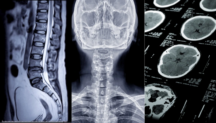
The choice depends on the patient’s symptoms, medical history, and the condition being investigated.
You can also learn more about CT scan services in Bhubaneswar.
What Is the Cost of a Spine MRI in Bhubaneswar?
The cost of a Spine MRI in Bhubaneswar varies depending on several factors. There is no single fixed price applicable to every diagnostic centre or type of examination.
Some important factors that influence the cost include:
- Spinal region: Cervical, thoracic, lumbar, and whole-spine MRI examinations may have different prices.
- Use of contrast: MRI scans performed with contrast material may cost more than routine non-contrast examinations.
- MRI equipment: Scanner strength and imaging technology can influence the overall cost.
- Diagnostic centre: Location, infrastructure, reporting services, and other facility-related factors may affect pricing.
- Imaging requirements: Specialised imaging protocols may involve additional charges.
Before scheduling your examination, confirm the scan type, whether contrast is required, and the total cost with the diagnostic centre.
For patients looking for aSpine MRI scan in Bhubaneswar, Prolife Diagnostics provides MRI services using 1.5T MRI technology, with imaging options including cervical, thoracic, dorsal, and lumbar spine MRI. Contact the centre for current availability and pricing.
When Should You Consult a Doctor?
Not every episode of back or neck pain requires an MRI. Many uncomplicated cases improve with appropriate medical management and physical activity.
However, you should consult a doctor if you experience:
- Persistent or worsening back or neck pain.
- Pain that spreads into the arms or legs.
- Recurrent numbness or tingling.
- Unexplained weakness in the arms or legs.
- Pain following an accident or significant injury.
- Symptoms that do not improve with recommended treatment.
Seek urgent medical attention if you experience new loss of bladder or bowel control, numbness around the groin or saddle area, or rapidly progressing leg weakness. These symptoms may indicate serious nerve compression and require immediate assessment.
Your doctor will determine whether a Spine MRI or another diagnostic test is necessary.
Related Articles: Learn More About MRI Scans
Explore these helpful resources from Prolife Diagnostics to understand MRI scans, their uses, and diagnostic services:
- Why Prolife Diagnostics Is the Best MRI Scan Center in Bhubaneswar
- What Medical Conditions Might Require an MRI Scan?
- What Common Diseases Can Be Diagnosed with an MRI Scan?
Get the Right Diagnostic Support at Prolife Diagnostics
Spinal pain and related neurological symptoms should not be ignored, particularly when they persist, worsen, or interfere with everyday activities. A Spine MRI can provide valuable information about the spinal cord, discs, nerves, and surrounding structures when imaging is clinically indicated.
At Prolife Diagnostics, Bhubaneswar, we offer advanced MRI imaging using 1.5T MRI technology to support the evaluation of different spinal conditions. Our MRI services include cervical, thoracic, dorsal, and lumbar spine examinations.
Whether your doctor recommends an MRI for persistent back pain, suspected disc problems, or nerve-related symptoms, choosing an appropriate diagnostic examination is an important step towards informed medical care.
Contact Prolife Diagnostics to enquire about Spine MRI availability, preparation instructions, and pricing in Bhubaneswar.